
Minimally Invasive Pain Management
Millions of people live with chronic back and neck pain, which can affect not only the back and neck, but other parts of the body like the hip, arms and legs and can contribute to general all-over body pain. For many people living with chronic pain, finding pain relief can be tough. A lot of trial and error is involved to find a pain treatment that works. Interventional pain management may help chronic pain patients cope with their pain.
Similar to other pain management treatments, such as taking prescription medications, interventional pain management can help you manage your pain. But what makes interventional pain management different is that it uses techniques, such as injections to directly address the source of your pain.
Below are some of the most commonly used interventional pain management techniques.
- Injections (also called nerve blocks)- work to provide temporary pain relief. They send powerful medications, such as steroids and opioids, onto or near your nerves to relieve pain.
- Epidural steroid injection- is an injection in your lumbar spine (low back). This injection sends steroids directly to the nerve root that’s inflamed.
Other common injections are facet joint injections, single nerve root blocks, and sacroiliac joint injections.
You’ll most likely need 2 or 3 injections for maximum benefits, but you should not have more than that due to the potential side effects related to steroid and other medications administered.

EMG (Electromyogram)
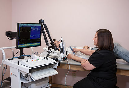
An electromyogram (EMG) is a test that is used to record the electrical activity of muscles. When muscles are active, they produce an electrical current. This current is usually proportional to the level of the muscle activity. An EMG is also referred to as a myogram.
EMGs can be used to detect abnormal electrical activity of muscle that can occur in many diseases and conditions, including muscular dystrophy, inflammation of muscles, pinched nerves, peripheral nerve damage (damage to nerves in the arms and legs), amyotrophic lateral sclerosis (ALS), myasthenia gravis, disc herniation, and others.
EEG (Electroencephalogram)
An electroencephalogram (EEG) is a test that measures and records the electrical activity of your brain. Special sensors (electrodes ) are attached to your head and hooked by wires to a computer. The computer records your brain's electrical activity on the screen or on paper as wavy lines. Certain conditions, such as seizures, can be seen by the changes in the normal pattern of the brain's electrical activity.
Vascular Imaging
Vascular imaging, also called ultrasound scanning or sonography, involves the use of a small transducer (probe) and ultrasound gel to expose the body to high-frequency sound waves. Ultrasound is safe and painless and produces pictures of the inside of the body using sound waves. Because ultrasound images are captured in real-time, they can show the structure and movement of the body's internal organs, as well as blood flowing through blood vessels.
A Doppler ultrasound study is usually part of a vascular ultrasound examination.
Doppler ultrasound is a special ultrasound technique that evaluates blood flow through a blood vessel, including the body's major arteries and veins in the abdomen, arms, legs and neck
Vascular Ultrasound is a useful way of evaluating the body's circulatory system. Vascular ultrasound is performed to:
-
help monitor the blood flow to organs and tissues throughout the body.
-
locate and identify blockages (stenosis) and abnormalities like plaque or emboli and help plan for their effective treatment.
-
detect blood clots (deep venous thrombosis (DVT) in the major veins of the legs or arms.
-
determine whether a patient is a good candidate for a procedure such as angioplasty.
-
evaluate the success of procedures that graft or bypass blood vessels.
-
determine if there is an enlarged artery (aneurysm).
-
determine the source and severity of varicose veins
